
Internship offer: Master's or Engineering Internship – 2025/2026 Medical imaging, Graphs, Artificial Intelligence
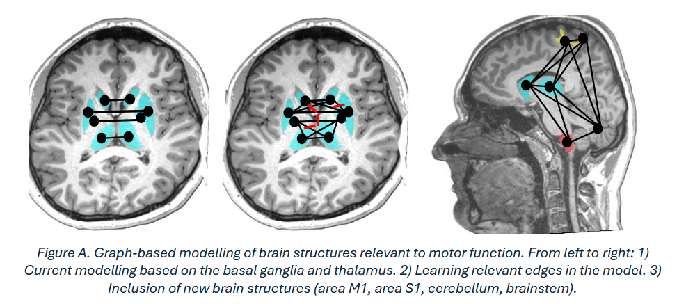
Around 30% of children who suffered a neonatal ischaemic stroke develop permanent motor disorders known as cerebral palsy (Chabrier, et al. 2019). Studying key regions of the brain may help to better understand the manual motor impairments caused by (Craig, Carlson and Kirton 2019) (Hassett, et al. 2022) (Ilves, et al. 2022). Understanding the phenomena resulting from the presence of this early brain lesion can rely on characterisation through imaging. Previously, we demonstrated a link between morphological information extracted from T1 morphometric MRIs and manual motor functions in children who suffered a neonatal stroke (Coupeau, et al. 2024). The proposed method is based on a graph neural network (GNN) (Bacciu, et al. 2020) designed to predict an individual's motor performance based on a graph of the brain. More specifically, each structure of interest in the brain is represented as a node incorporating morphometric properties derived from T1 MRI (Figure A-1). The nodes are connected by edges providing information on the spatial relationships between structures. The promising results obtained still need improvement, particularly for predicting fine motor skills.
A first way to improve the model is to enrich the spatial relationships embedded in the graph edges and to automate the elimination of irrelevant edges in the GNN (Figure A-2) as recently proposed (Zheng, et al. 2020) (Sui, et al. 2024). The current work is based on a 2-to-2 connection between structures (Figure A-1) based on a priori. The challenge is to tackle a classic problem in graph data science, relating to learning the relevant topology of a graph to perform a specific task. A second option being considered is to enrich the graph by adding new nodes associated with other brain structures involved in the motor system (Figure A-3). In addition to the basal ganglia and thalamus currently considered, we wish to consider the primary motor area (movement execution), the cerebellum (movement coordination and adjustment) and the brainstem (transmission of motor impulses to the muscles).
The aim of the internship is to implement the two areas for improvement described above and to study their impact on predicting motor function after a neonatal stroke.
The internship will begin with an exploration of the team's Python codes (Coupeau, et al. 2024). In a second stage, the intern will be tasked with adapting the GNN model to learn how to eliminate 2 edges that are irrelevant to the prediction. The aim is to move away from working with graphs connected in pairs and instead provide the network with a model without any a priori. For this part, the intern will work with a PhD student from the team. Programming will be done using the PyTorch geometric library (Fey et Lenssen 2019). The final task will be to enhance the modelling by adding nodes associated with new brain structures. The structures of interest have already been segmented by the team on the T1 MRIs; all that remains is to extract their morphometric properties.
During the internship, the intern will be invited to participate in monthly research meetings with medical researchers at Angers University Hospital. If the results are conclusive, they may be presented in a journal article or a conference paper.
The internship will take place at the LARIS laboratory for a period of 5-6 months starting in February 2026.
The intern will receive a stipend of €4.35 per hour of actual attendance. The University of Angers may cover 75% of the cost of your transport pass, upon presentation of proof of purchase, up to a limit of €101.75 per month.
Contact :
Patty Coupeau (patty.coupeau@univ-angers.fr)
To apply for this internship, please provide:
- CV + cover letter
- Master's 1 transcripts
- Letter of recommendation (optional)
Références :
- Bacciu, Davide, Federico Errica, Alessio Micheli, and Marco Podda. "A gentle introduction to deep learning for graphs." Neural Networks 129 (2020): 203-221.
- Chabrier, Stéphane, et al. "From congenial paralysis to post-early brain injury developmental condition: Where does cerebral palsy actually stand?" Annals of Physical and Rehabilitation Medicine 63, no. 5 (2019): 431-438.
- Coupeau, Patty, Josselin Démas, Jean-Baptiste Fasquel, Lucie Hertz-Pannier, Stéphane Chabrier, et Mickael Dinomais. «Hand function after neonatal stroke: a graph model based on basal ganglia and thalami structure.» NeuroImage: Clinical 41 (2024): 103568.
- Craig, Brandon T, Helen L Carlson, and Adam Kirton. "Thalamic diaschisis following perinatal stroke is associated with clinical disability." Neuroimage Clinical 21 (2019): 101660.
- Fey, Matthias, et Jan E Lenssen. «Fast Graph Representation Learning with PyTorch Geometric.» ICLR Workshop on Representation Learning on Graphs and Manifolds, 2019.
- Hassett, Jordan, Helen Carlson, Ali Babwani, and Adam Kirton. "Bihemispheric developmental alterations in basal ganglia volumes following unilateral perinatal stroke." Neuroimage Clinical 35 (2022): 103143.
- Ilves, Nigul, et al. "Ipsilesional volume loss of basal ganglia and thalamus is associated with poor hand function after ischemic perinatal stroke." BMC Neurology 22, no. 1 (2022): 23.
- Sui, Yongduo, Xiang Wang, Tianlong Chen, Meng Wang, Xiang He, et Tat-Seng Chua. «Inductive Lottery Ticket Learning for Graph Neural Networks.» Journal of Computational Science and Technology, 2024.
- Zheng, Cheng, et al. «Robust Graph Representation Learning via Neural Sparsification.» International Conference on Machine Learning, 2020.
