- Index
- >Projects
- >Past projects
- >PROCHIP
PROCHIP Research Project
Chromatin organization PROfiling with high-throughput super-resolution microscopy
Group : Information, Signal, Image and Life Sciences
Labelling: none
Duration: 3 years (2018 - 2021)
Funding: EU H2020 FET Open program
Staff involved from LARIS: David Rousseau, Ali Ahmad
Project partners: 6 partners in 3 European countries
What is the PROCHIP research project?
![]()
The goal of the project is to build a high-throughput super-resolution microscope in a microfluidic chip smaller than a coin capable of providing high resolution imaging of hundreds of cells at the diffraction limit and beyond, with minimal photo-toxicity.
Target
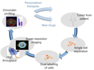
The targeted breakthrough is to interrogate cancer heterogeneity at single cell level by classifying freshly isolated cancer samples based on their chromatin architecture: we aim to use chromatin alteration as a marker for cancer.
Vision
Our long term vision is to develop a new enabling technology, able to extract cancer cell from a patient, assess alterations in chromatin architecture at single cell level, and define a targeted personalized therapy or drive the development of new drugs and pharmaceutical treatments.
Consortium
PROCHIP Project has been granted a 2,5€ million from the EU H2020 FET Open programme. The 3-years project will be carried out by an international consortium of 6 organizations from 3 European countries, led by the Italian National Research Council.
![]()
![]()
![]()

![]()
![]()
Project overview
Metastasis is the leading cause of cancer death among cancer patients. Cancer genomics showed that tumor progression to metastasis formation is poorly supported by further genetic alterations, implying that the adaptation capacity of disseminating tumor cells to foreign microenvironments rely on epigenetic alterations. Epigenomic reprogramming plays a central role in cancer progression and metastasis formation, supporting tumour heterogeneity, which represents a challenge for precise diagnosis and targeted therapy.
To decipher the pathogenic role of tumor heterogeneity in tumor progression and therapeutic response, it is crucial to define epigenetic alterations, at the single cell level, within the cancer cell populations.
The aim of the project is therefore to syudy cancer heterogeneity at single cell level by classifying freshly isolated cancer samples based on their chromatin architecture: we aim to use chromatin alteration as a marker for cancer. In order to achieve this result we will develop a superresolution microscope with high throughput capabilities, able to acquire thousands or even tens of thousands samples. Extracting and imaging large number of samples will allow performing statistically relevant biological studies that take into account the variability of cellular behaviour in the same pathological context, and creating highly specific phenotyping procedures required for precision medicine.
Ideally, microscopy devices should have a flexible design that allows customization for various samples and should be low-cost for a widespread diffusion in the context of personalized medicine. To fulfill these requirements we will develop a lattice light sheet microscope using lab-on-chip technologies: the illumination path and sample delivery systems will be fully integrated in a mm-scaled lab-on-chip. Using this device we aim to provide high-resolution imaging of cells and sub-cellular structures as chromatin network at the diffraction limit and beyond, with minimal photo-toxicity and to generate high-throughput data by scanning them in automatic fashion within a fluidic network.
The long term vision of PROCHIP project is to develop a new enabling technology, able to extract cancer cell from a patient, assess alterations in chromatin architecture at single cell level, and define a targeted personalized therapy or drive the development of new drugs and pharmaceutical treatments.
In details the objectives of the project are to:
1) develop a miniaturized three-dimensional fluidic network integrated on a glass chip and its relative pumping system for automatic sample scanning under a microscope.
2) develop a super-resolution microscope, whose illumination and sample scanning components are integrated in a lab-on-chip (super-resolution microscope on-chip).
3) develop the protocols and the technology for isolation of primary and metastatic tumor cells suitable for high-throughput imaging and data analysis at superresolution based on the image simulator.
4) assess cancer cell heterogeneity by querying chromatin domains as functional biomarkers.
Website: https://pro-chip.eu/
MicroVIP: Microscopy simulation software on VIP https://www.creatis.insa-lyon.fr/site7/en/MicroVIP